Introduction
The World Health Organization (WHO) has defined probiotics as ‘living organisms’. The function of probiotics has been studied for antibiotic-related diarrhea, improvement of hypertension, reduction of blood cholesterol level, intestinal infection caused by pathogenic bacteria, improvement of gastrointestinal diseases and improvement of blood lipid status [1–6]. The probiotic categories used today include various yeast species, including Saccharomyces bouladii, and bacteria such as lactic acid bacteria (LAB) [7]. LAB are important microorganisms that have long been used in modern industrial and traditional food fermentations. In addition to adding flavor, many foods fermented by LAB are believed to add health benefits [8]. Due to the increasing benefits of using LAB, the use of LAB in food is increasing in recent years.
Kombucha is commonly known as ‘tea fungus’ by fermenting sugar-sweetened black tea for 10–12 days at room temperature [9]. Kombucha tea is a beverage obtained using the fermentation of black or green tea, usually black tea, by a symbiotic culture of acetic acid bacteria (AAB) and yeast [10]. Yeast cells hydrolyze sucrose to glucose and fructose to produce ethanol, while AAB convert glucose to gluconic acid and fructose to acetic acid [11,12]. This is the main metabolic pathway in Kombucha. The main metabolites identified in fermented beverages are acetic, lactic, gluconic and glucuronic acids, as well as alcoholic ethanol and glycerol [13–15]. Organic acids produced during fermentation prevent rancidity of beverages from contamination by unwanted microorganisms and bacteria introduced from outside [13,16]. Kombucha also showed antibacterial activity [16]. The antimicrobial activity of Kombucha is measured according to its acetic acid content [17]. Accordingly, recent studies on Kombucha have demonstrated that the antimicrobial activity against pathogenic microorganisms is mainly due to acetic acid [16,18]. One report investigated the interaction of growth rates, biomass and secondary metabolites between the LAB in kefir milk, a fermented milk beverage made using grain as a yeast and bacterial fermentation starter, and the AAB in Kombucha. LAB have been shown to improve the survival of AAB [19]. In addition, the vitamin B complex secreted by AAB provides a favorable environment for the growth of other yeasts and LAB in kefir grains [20]. Therefore, in this study, the importance of LAB in Kombucha fermentation was evaluated, and biological functions such as organic acid production, anti-inflammatory, antibacterial activity, and antioxidant activity of kelp were evaluated according to whether or not LAB were inoculated. Also, instead of using green tea and black tea, which are commonly used as raw materials for Kombucha, the experiment was conducted using green tea extract as a substrate. Therefore, Kombucha that was not inoculated with LAB was named Kombucha without Lactic acid bacteria (KWOL), and Kombucha that was inoculated with LAB was named Kombucha with Lactic acid bacteria (KWL).
Materials and Methods
After solubilizing sucrose in 70 g/L of deionized water and boiling, green beans were added at 5 g/L and extracted at 90°C for 30 min. Then, the mixture was filtered using a 300 mesh sterilizer and cooled to room temperature. After mixing the strain (Lactobacillus paracasei DK215, Saccharomyces cerevisiae C3, and Acetobacter pasteurianus P2) diluted to 1×10³ CFU/mL in the sterilized solution, the inlet was sealed using sterile gauze, and aerobic fermentation and stationary fermentation were performed at 30°C.
Organic acids (acetic, citric, formic, lactic, and glucuronic acid) were measured using HPLC apparatus. Samples were prepared by centrifugation at 12,000×g and 4°C for 30 min, and were then filtrated with a 0.45 μm sterile syringe membrane filter (SIGL). The filtrate was used as a sample for injection into the HPLC injector. 10 μm of the filtered sample was injected was subjected to high-performance liquid chromatography (Waters, USA) on Hydrosphere C8 column (250×4.6 mm, particle size 5 μm, YMC, Japan) using 20 mM NaH2PO4 (pH 2.8). The flow rate maintained at 1.0 mL/min and the temperature of 30°C. The concentration of organic acids was measured by comparing the corresponding peak area against the standard curve and multiplied by the dilution factors. All samples were analyzed in triplicate (Table 1).
The antimicrobial activity of Kombucha tea samples was assessed by agar diffusion assay [21] against four human pathogenic bacteria Gram-positive Staphylococcus aureus (KCTC3881, Korea) and Gram-positive bacilli Bacillus cereus (KCTC362A) and Gram-negative bacilli including E. coli (KCTC1682), Salmonella enterica (KCTC2054). TSA Agar (20 mL) was poured into each Petri dish (90 mm diameter). Suspensions (100 μL) of target strain cultured for 24 h were spread on the plates uniformly, and wells of 9 mm diameter were made with a sterile cork borer. Kombucha tea samples were centrifuged at 8,000×g for 15 min at 4°C and filtration through a PVDF 0.45 μm SIGL to remove cell debris. For the purpose of control and comparison, acetic acid samples at the same concentration as that of fermented tea after 7 days (7.6 g/L) were prepared. In the same way, pH 4 and 7 samples of unfermented and fermented tea were obtained by adjusting the pH with 1 M HCl or 1 M NaOH. Heat-denatured samples were treated at 100°C for 5 min. Samples (100 μL) were then transferred into the wells of agar plates inoculated with target strains. The plates were first put at 4°C for 2 h to make a prediffusion of tea sample into the agar. The plates were then incubated at 30°C. The diameter of the inhibition zone was measured after 12–15 h.
RAW264.7 (mouse macrophage cell) were obtained from the KCTC (Jeongeup, Korea). RAW 264.7 cells were cultured in Dulbecco’s modification of Eagle’s medium (DMEM), supplemented with heat-inactivated 10% FBS at 37°C, under an atmosphere of 5% CO2. After centrifuging at 10,000×g for 5 min at 4°C the growth medium and discarding, the medium was changed every two day. To evaluate the protection of cells, cultured RAW264.7 cells were seeded in 96-well plates (100 μL per well, for cell viability determination and nitric oxide production inhibition) at a loading concentration of 1×10⁵ cells/mL.
Cell viability was determined by MTT assay as previously reported [22,23]. RAW264.7 cells were seeded in 96-well cell culture plates at density of 1×10⁵ cells/well and incubated for 16–18 h. After discarding the growth medium, cells were treated with Kombucha samples diluted with various concentrations of acetic acid in serum-free medium for 20–22 h. After treatment, 100 uL of a 5 mg/mL 3-(4,5-dimethylthiazol-2-yl) -2,5-diphenyltetrazolium bromide (Sigma-Aldrich, USA) solution was added to each well (excluding blank wells), and the samples were re-incubated for 2–3 h in the dark in an incubator (37°C, 5% CO2). Culture supernatants were removed, and 100 uL Dimethyl sulfoxide (DMSO; Sigma-Aldrich) was added to each wells to completely dissolve formazan crystals. The absorption was measured at 540 nm [24].
The amounts of NO were calculated by measuring the amount of nitrite oxidized product [25]. RAW264.7 cells were seeded in 96-well cell culture plates at density of 1×10⁵ cells/well and incubated for 16–18 h. After discarding the growth medium, cells were treated with Kombucha samples diluted with various concentrations of acetic acid in serum-free medium for 2–3 h. Thereafter, lipopolysaccharide (LPS) (Sigma-Aldrich) at a final concentration of 100 ng/mL was treated and stimulated in the same volume in medium for 20–22 h. Then, 60 uL of the reaction mixture was withdrawn and added to a 96-well microplate, followed by 100 uL of Griess reagent (1% sulfanilamide, 0.1% N-(1-naphthyl)-ethylin diamine hydrochloride, 2.5% H3PO4). The absorbance at 540 nm was determined in a Microplate reader after 15 min at room temperature.
RAW264.7 cells were seeded in 96-well cell culture plates for 24 h. After removing the medium, RAW264.7 cells were treated with various concentrations of fraction samples in a serum free medium for 2–3 h. After that, LPS at a final concentration of 100 ng/mL was treated and stimulated in the same volume in serum free medium for 20 h. Cell-free supernatants were collected and the cytokine content was measured by a Mouse IL-1, IL-6 and Mouse TNF-α ELISA kit (Komabiotech, Korea) using an enzyme-linked immunosorbent assay (ELISA). The optical density of the microplate was read at 450 nm.
Results
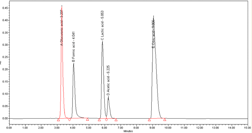
The concentration of Kombucha organic acid according to fermentation time was measured using HPLC. It was confirmed that the acetic acid concentration of Kombucha inoculated with LAB gradually increased to reach a maximum of 7.6 g/L on the 7th day of fermentation, and it was confirmed that glucuronic acid also reached 7 g/L on the 7th day of fermentation. However, in the case of Kombucha that was not inoculated with LAB, acetic acid was 3.1 g/L and glucuronic acid was 1.1 g/L, which was relatively low. The three organic acids except acetic acid and glucuronic acid were very little or not measured in both types of Kombucha studied (Table 2, Figs. 1-3).


In this study, the antibacterial activity of Kombucha against various pathogens was investigated using the agar diffusion method. The results are shown in Table 4. Acetic acid (7.6 g/L), unfermented tea adjusted to pH 5–7, and heat denatured (100°C, 5 min) tea were used as controls. Kombucha has been found to exhibit antimicrobial activity in S. aureus, E. coli, and S. enterica, except for B. cereus. Unfermented tea had no antibacterial activity against the target microorganism. Both Kombucha samples showed the highest level of antibacterial activity in E. coli, and relatively higher antibacterial activity was confirmed in Kombucha inoculated with LAB (Table 3 and 4).
The diameters of the clear zone of inhibition were as follows: –, no inhibition; +, 10≤diameter≤15; ++, 15≤diameter≤20; +++, 20≤diameter≤25 (mm); Acetic acid, acetic acid concentration of Kombucha (7.6 g/L); Unfermented tea, 1 M HCl or 1 M NaOH was used to adjust their pH; Heat-denatured: 100°C, 5 min; Natural pH: pH value of the sample without any adjustment.
The diameters of the clear zone of inhibition were as follows: –, no inhibition; +, 10≤diameter≤15; ++, 15≤diameter≤20; +++, 20≤diameter≤25 (mm); Acetic acid, acetic acid concentration of Kombucha (7.6 g/L); Unfermented tea, 1 M HCl or 1 M NaOH was used to adjust their pH; Heat-denatured: 100°C, 5 min; Natural pH: pH value of the sample without any adjustment.
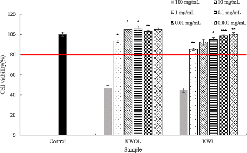
Cytotoxicity testing via MTT assay is widely used in in vitro toxicity studies. The cytotoxic MTT assay of mouse macrophage RAW264.7 cells confirmed the cell viability of 80% or more in the remaining samples except for the acetic acid concentration of 100 mg/mL in the Kombucha sample at a concentration of 1×10⁵ cells/well in 96 wells. Therefore, it was confirmed that both Kombucha samples were not cytotoxic in macrophages (FIg. 4).

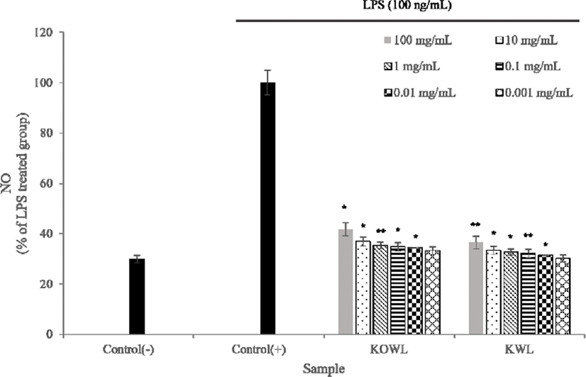
Excess NO production leads to serious diseases such as septic shock, stroke, arthritis, chronic inflammatory disease, and autoimmune diseases. As a result of the study, it was shown that NO production was suppressed as the concentration of the two Kombucha samples increased. Accordingly, it was shown that the inhibition of NO production during Kombucha fermentation was not induced by cytotoxicity, and it was confirmed that the NO production of Kombucha inoculated with LAB was relatively highly inhibited (FIg. 5).

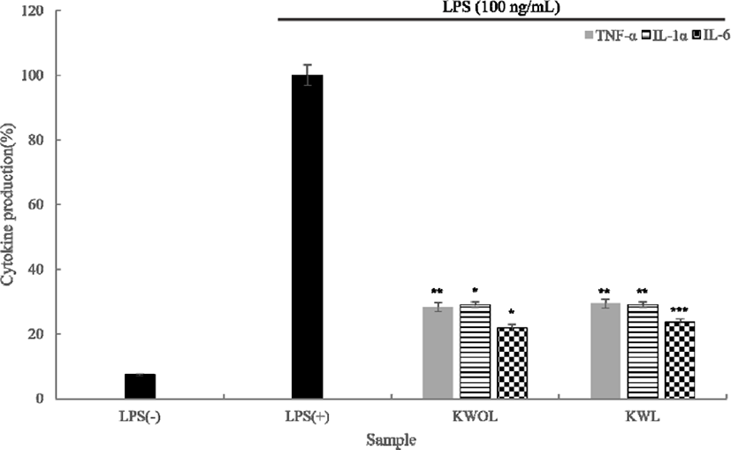
Cellular TNF-α, IL-6, and IL-1α levels were also assessed using a TNF-α, IL-6, and IL-1α specific ELISA kit. As a result of cytokine measurement using ELISA, the production of three cytokines (IL-1α, IL-6, TNF-α) was significantly lower than that of the LPS (+) group. FIg. 6. showed that LPS significantly stimulated TNF-α, IL-6, and IL-1α production. These results are believed to reduce the inflammatory response (FIg. 6).

Discussion
This study was conducted to evaluate the potentially beneficial biological functions of Kombucha inoculated with LAB, such as organic acid production, anti-inflammatory, antibacterial activity and antioxidant capacity, compared to Kombucha normally fermented using yeast and AAB. Lactobacillus paracasei DK215, Saccharomyces cerevisiae C2, Acetobacter pasteurianus P2 was selected and used in the experiment.
Glucuronic acid is the most important metabolite during Kombucha fermentation, improving immune response, anticancer, gastric ulcer treatment and antidiabetic are effective [26–29]. Therefore, in this experiment, as a result of measuring the content of a total of five organic acids including glucuronic acid at 3-day intervals during Kombucha fermentation, it was confirmed that 7.6 g/L of acetic acid on the 7th day of fermentation was reached in KWL, and Glucuronic acid was also It was confirmed that it reached 7 g/L. In the case of KWOL, the organic acid content was relatively low at 3.1 g/L for acetic acid and 1.1 g/L for glucuronic acid. Except for acetic acid and glucuronic acid, the three organic acids were very little or not measured in the two types of Kombucha studied. This was judged to improve the fermentation characteristics in symbiosis with other strains when inoculating LAB into Kombucha.
A previous study reported that the antimicrobial activity of Kombucha was determined by the acetic acid content [16]. The acetic acid content at the end of fermentation was set as a control. In addition to S. aureus and E. coli, which are strains commonly used in food, in this experiment, B. cereus and S. enterica were added and compared with various control groups. As a result of the experiment, it was confirmed that it exhibits antibacterial activity in S. aureus, E. coli, and S. enterica except for B. cereus. Unfermented tea had no antibacterial activity against target microorganisms, but both Kombucha samples showed the highest level of antimicrobial activity in E. coli, and relatively higher antimicrobial activity was confirmed in KWL.
After culturing RAW264.7 cells, which are mouse macrophages, in 96 wells at a level of 1×10⁵ cells per well, the cytotoxicity MTT assay results of samples diluted by acetic acid concentration showed cytotoxicity or growth at the remaining concentrations except for 100 mg/mL. No facilitation action could be confirmed. Afterwards, it could be confirmed based on 80% or more, which is the reference point for cell viability in the MTT assay. LPS is a lipopolysaccharide, a structure that exists on the surface of bacteria, and is a substance that can be used as an antigen to induce an immune response in the host. NO is a substance produced by such an immune response, and when looking at the amount of production, it was confirmed that the Kombucha fermented sample had a lower NO production than the LPS-treated group. In addition, compared with KWOL, it was confirmed that the value of the KWL sample was close to that of the LPS untreated group. Cytokine (IL-1α, IL-6, TNF-α) production was significantly lower than that of the LPS (+) group, and based on this result, it was possible to find the possibility of anti-inflammatory activity in the Kombucha sample.
Therefore, in this study, it was judged that inoculation of Lactobacillus paracasei DK215 improved the antibacterial and the change of glucuronic acid content during fermentation, compared to the original Kombucha using only yeast and AAB. It has been confirmed that the improvement of the biological function of Kombucha inoculated with these LAB can once again prove the value of the LAB in the fermented food and beverage industry. However, further investigation into the mechanisms involved in the fermented beverage raw material substrates involved in the production of biological metabolites is deemed necessary.
